Normal S1 and S2 at normal speed and at 60% normal speed, +25% pitch
What if we wanted to auscultate a normal S1/S2 but at the base positions, such as the Aortic area?
 Aortic, Bell, Patient Sitting
Normal S1 and S2 stethoscope is at aortic position, still using the bell. Note the louder S2 compared with Normal S1/S2 at Apex
Aortic, Bell, Patient Sitting
Normal S1 and S2 stethoscope is at aortic position, still using the bell. Note the louder S2 compared with Normal S1/S2 at Apex
(no image available)
Splitting of S2, Normal
Pulmonic, Bell, Supine
Physiologic Splitting (4 cardiac cyles, looped total of 3x); 2nd beat is at peak inspiration and has best split of S2, 4th at peak expiration
Same as above, but slowed to 60% and only 2 loops
Part 2
Non-Physiologic S2 Split
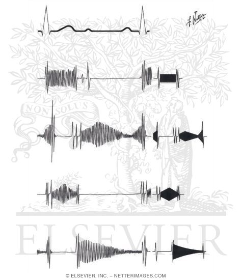
Use the image from Figure 11-35 to demonstrate difference in splitting
Wide Splitting in this audio is caused by a Right Bundle Branch block;
note that split is easily appreciated, despite respiratory variation
S3, third heart sound
 Apex, Bell, Left lateral decubitus
Listen to the S3 at normal speed and pitch
Here is the same audio of the S3 at normal speed but pitch changed to better appreciate sound
Finally this is the S3 with 60% the original speed and higher pitch
Apex, Bell, Left lateral decubitus
Listen to the S3 at normal speed and pitch
Here is the same audio of the S3 at normal speed but pitch changed to better appreciate sound
Finally this is the S3 with 60% the original speed and higher pitch
S4, fourth heart sound
 Apex, Bell, Left lateral decubitus
Listen to an S4 at Normal speed, left lateral decubitus, apex, bell
Same audio sample of S4 but slowed to 60%
Apex, Bell, Left lateral decubitus
Listen to an S4 at Normal speed, left lateral decubitus, apex, bell
Same audio sample of S4 but slowed to 60%
Part 3
Students, below are link to four unique heart sounds. In an effort to make things fair,
the sounds are mixed up depending on the day of the week your physical diagnosis class meets.
You will be assigned one of these 4 sounds. Your responsibility is to identify the abnormality
and demonstrate to the class the following:
- Correctly identify their audio sample’s disease (also highlight if it is a systolic or diastolic murmur)
- Playback audio and defend your answer choice
- Demonstrate how best to auscultate this finding
- Provide location of best auscultation, position
- Provide areas to auscultate for radiation; if none then make note of this
- On an audiogram, what would the shape of this murmur be
- Any associated signs (e.g. ejection click, narrow pulse pressure, etc.)
- If relevant, demonstrate where to auscultate for any radiation
- Outline symptoms of the disease (can separate by acute vs chronic)
- Outline any relevant diseases associated with your valvular disease
Reference Chart

By Madhero88 - Own workReference netter image, CC BY-SA 3.0, https://commons.wikimedia.org/w/index.php?curid=9663180L
